A horse’s foot is a funny thing, and not the good Billy-Crystal-hosting-the-Oscars kind of funny
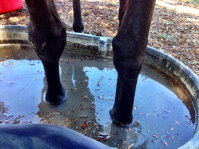
Photo by Gina Spadafori
Cooling a horse's feet in a slurry of ice and water immediately following a known laminitis trigger, such as over-eating, can reduce the incidence of clinical laminitis.
The second leading cause of death in horses in the United States is a condition called laminitis. (Colic is the leading cause.) This equine arch-nemesis also uses the alias “founder.”
Laminitis, an inflammation of the connective tissues inside the black hole of the equine foot, made headlines a while back after the breakdown and eventual euthanasia of the racehorse Barbaro.
A horse’s foot is a funny thing, and not the good Billy-Crystal-hosting-the-Oscars kind of funny. We’re talking more along the lines of high-tech equipment that functions wonderfully as long as you don’t breathe on it wrong or look at it strangely.
Anyone who has ever been on the receiving end of a horse’s foot might have a hard time believing it, but inside that hard, seemingly-indestructible, and occasionally destructive hoof is a complex and delicate apparatus.
If you cut a horse’s foot in half – please do not try this at home – you will see a series of bones held in place by tendons, ligaments, and funny looking ruffles that look like some weird sea anemone. You will also find a literal net of blood vessels that wraps around the inside of the foot. (This is a gross oversimplification in order to get a textbook chapter’s worth of description into one paragraph before most readers fall asleep or get distracted by the pile of dishes in the sink.) The ruffly, leafy-looking connective tissue is the laminae (also called lamellae.)
These laminae help hold the third bone of the phalanx, a triangular-shaped thing also called (somewhat ominously) the coffin bone, in place. The tendons that attach to the coffin bone from behind pull back and upward. The laminae connect the coffin bone to the inside of the hoof capsule around the sides and front, preventing the tendons from jerking it out of place. This nice balance also prevents that net of blood vessels mentioned earlier from getting squished or otherwise disrupted, thus maintaining the foot in a state of circulatory bliss.
However, when this balance gets disrupted, things go awry.
By now you know the “laminae” part of laminits. So, let’s get to the –itis. In medicalese (well, Latin), “itis” means “inflammation.” Laminitis=inflammation of the laminae.
“What causes laminitis?” you ask. That, folks, is the $64 million question. While we know a long list of specific triggers for laminitis, the exact pathways in the body between cause and outcome are still a little murky and very, very complicated. The research on this disease has come a long way even in recent years, but right now, it’s the known triggers that probably matter most to horse owners.
They can be broken down into a few categories:
- Ingested/Metabolic – “Grain overload” is one of the most common causes of laminitis. A more accurate term would be “sugar overload.” When a horse eats more high-carbohydrate feed than his body can handle, the resulting metabolic meltdown often directs the full force of its fury at the feet. For horses with normal metabolism, an overload event would be something like breaking into the feed shed and treating the grain barrels as a Las Vegas buffet. But for horses with more delicate constitutions or what is known as equine metabolic syndrome, a short picnic on some lush grass can tip the system over the edge.
Black walnut shavings also cause laminitis, though the jury is still out on whether horses have to eat the shavings or if simple exposure will do it.
- Infectious/Inflammatory – Anything that triggers a significant inflammatory reaction in a horse can potentially lead to laminitis. This might include infectious causes such as a virus, gastrointestinal infection, uterine infection, or retained placenta after foaling. It could also include severe reactions to things like drugs, snakebite, or vaccines.
- Impact – By impact, I really mean anything that alters or increases the forces exerted on the foot. This could be an improper hoof trim, increased weight bearing on one foot because the opposite foot is injured, or concussive work on a hard surface.
- Idiopathic – Idiopathic is the fancy catch-all term for “Heck if we know.” This is the island of unknown causes, falling loosely into the realm of “stuff happens."
Where a cause can be determined, treating or removing the cause is a BIG deal. Treat the infection, get the horse off the pasture, treat all horses that get into the feed room BEFORE there are any signs of illness. The best treatment for laminitis is to try to interrupt the source before it can start.
But that’s not always possible, and sometimes events spiral into the underworld of lamellar inflammation.
When the laminae become inflamed, several things happen. As you might guess, none of them are super fun.
- Pain – If you’ve ever had an ingrown toenail or an infection under your fingernail, you might have a faint idea of laminitic pain – if you were bearing roughly 350 pounds on that one digit, and a similar amount of weight on the equally painful digit on the other side. Now, realize that you are standing in that pain constantly. Horses don’t have Lazy-Boy chairs and they are physiologically and psychologically unable to spend much time lying down.
- Edema – When a tissue becomes inflamed, fluid begins to accumulate in that tissue, causing swelling. This fluid is referred to as edema. In most soft tissues, you see an external swelling and possibly redness (think of a sore, swollen arm after a bee sting). Since the horse’s foot is enclosed by a rigid capsule (the hoof wall), there is nowhere for the tissue to swell. This trapped swelling contributes to the pain (see above) and instability in the foot (see below).
- Circulatory disruption – Consistent blood flow depends on blood volume, pressure, thickness of the blood, and vessel diameter. When these things change, flow changes. In laminitis, the blood vessel diameter can narrow due to pain and physical constriction as the foot loses structural stability (see below). Inflammation and dehydration (when your feet hurt, you tend not to walk to water) can both decrease the blood pressure and increase the sludginess of the blood. To summarize: sludgy blood moving at lower pressure through smaller tubes = not good for circulation. When the circulation is compromised, the tissues don’t get the oxygen and nutrients they need. And tissues starved for nutrients and oxygen die. This contributes to…
- Instability of the foot – This is the biggest problem associated with laminitis and the primary reason that laminitis is such a disastrous disease. Remember how we said that the laminae hold the coffin bone in place and counteract the pull of the tendons? Well, when those laminae become inflamed and filled with fluid, they lose their connections to each other, sort of like Velcro losing its fuzz.
Since the tendons don’t stop pulling, the forces involved disrupt the position of the coffin bone within the foot, causing it to pull away from its attachments and either sink downward or to rotate, in some cases severely enough that the tip of the bone penetrates the sole of the foot. When that happens, the bone is open to infection, and the fragile net of blood vessels has been so disrupted that tissues begin to die of starvation – a process called necrosis.
There’s a whole lot of potential bad and not any good in any case of laminitis, but not every case ends in apocalyptic despair. While there is no one “cure” for laminitis, early intervention and teamwork by veterinarian, farrier, and horse owner can go a long way toward minimizing the damage.
Treatment is four-pronged:
- Remove or treat the cause – see above
- Treat the pain – usually with medication
- Support the foot – deep bedding and special shoeing/pad arrangements are used to make sure the pressure on the foot is dispersed evenly
- Trim the foot back into the appropriate balance – this requires X-rays and GREAT communication between veterinarian and farrier.
None of these things can be accomplished without every member on the horse’s “team” being completely on board with the program.